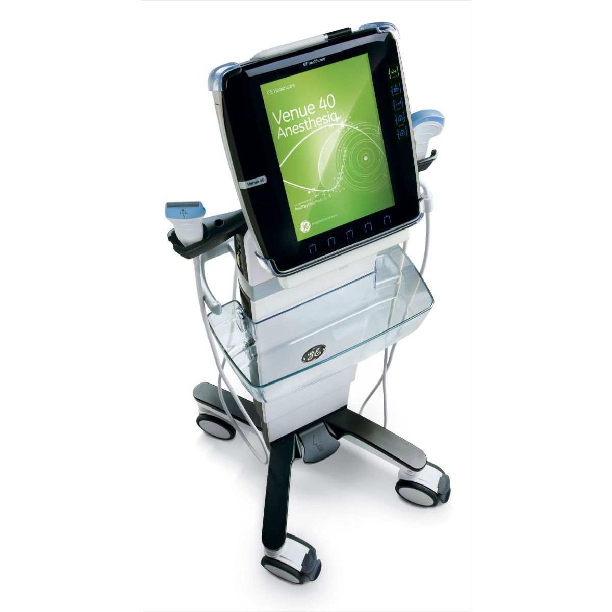
GE VENUE 40 Ultrasound (Portable) stands at the forefront of portable medical imaging technology, offering high-resolution imaging capabilities and exceptional versatility in diverse clinical environments.
Key Features and Functionality:
1. Portability and Mobility: The defining feature of the GE VENUE 40 Ultrasound (Portable) is its portability. With its compact size and lightweight build, healthcare providers can effortlessly transport it to various locations.
2. Image Quality Excellence: Despite its portable nature, the VENUE 40 delivers exceptional image quality. Armed with cutting-edge transducer technology and advanced imaging algorithms, it enables healthcare professionals to visualize organs.
3. Point-of-Care Versatility: The system’s portable design and user-friendly interface make it well-suited for point-of-care diagnostics. Physicians can conduct ultrasound examinations directly at the patient’s bedside, facilitating immediate assessments and prompt medical interventions.
4. Real-Time Imaging: Real-time imaging capabilities allow healthcare providers to observe dynamic changes within the body. This feature is crucial for monitoring procedures, guiding interventions, and assessing treatment effectiveness.
5. User-Friendly Interface: GE VENUE 40 Ultrasound (Portable) boasts an intuitive interface that simplifies operation. Accessible imaging modes and settings streamline the scanning process, enabling clinicians to efficiently navigate options and optimize imaging parameters.
Clinical Applications:
1. Point-of-Care Diagnostics: The GE VENUE 40 Ultrasound (Portable) portability empowers healthcare providers to perform immediate and accurate diagnostics at the bedside. This is particularly advantageous in critical care settings and during emergencies.
2. Emergency Medicine: In situations where rapid diagnostics are vital, the VENUE 40 excels. Its swift boot-up time, high-quality imaging, and portability enable healthcare professionals to promptly assess patients and make informed treatment decisions.
3. Obstetrics and Gynecology: The system’s image quality is especially beneficial for visualizing fetal development, assessing reproductive health, and diagnosing conditions in obstetrics and gynecology.
4. Musculoskeletal Imaging: The VENUE 40’s high-resolution imaging capabilities are essential for evaluating musculoskeletal structures. Physicians can diagnose injuries, assess joint health, and examine muscles and tendons with accuracy.
Maintenance and Quality Assurance:
To uphold the performance of the GE VENUE 40 Ultrasound, routine maintenance is crucial. Regular servicing, calibration, and software updates ensure consistent image quality and optimal functionality.
The GE VENUE 40 Ultrasound exemplifies the transformative impact of portable medical imaging technology. Its ability to deliver high-resolution imaging, real-time diagnostics.
GE Venue 40 Specifications
Dimensions
- Height: 11.1” (28.2 cm)
- Width: 10.8” (27.4 cm)
- Depth: 2.1” (5.4 cm)
- Weight: 8 lbs (3.6 kg)
Power
- Voltage: 100-240 V AC
- Frequency: 50/60 Hz
- Power: Max. 180 VA
Console Design
- Tablet Style
- Lithium-Ion Battery Pack (standard)
- 1 transducer port with SC-connector
- Speaker
- Docking station and docking cart are options
- Stylus
Display
- Size: 10.4-inch High-Resolution Color LCD
- Resolution: 800 x 600
- View: 170-degree wide angle view
Packages
- Musculoskeletal
- Vascular Access
- Anesthesia
- Point of Care
- Interventional
Transducer Types
- Linear Array
- Phased Array
- Convex Array
Operating Modes
- B-Mode
- M-Mode
- Color Flow Mode (CFM)
- Power Doppler Imaging (PDI)
- B-Steer + Needle Recognition
Software Features
- Applications
- Color/PDI
- M-Mode
- DICOM
- B-Steer + Needle Recognition
Hardware Options
- Battery Pack
- Docking Cart
- Docking Station
- 3-probe port
- Transducers
Media and Peripherals
- USB thermal B&W printer: Sony UPD-897 (option)
- Memory Stick (option)
- Footswitch (option)
B-Mode
- B Acoustic Output: preset in 3 steps, toggle for Low, Med, High
- Thermal Index: TI
- Gain: preset in 3 steps, toggle for Low, Med, High
- Harmonics: defined by the application
- Depth: 0.5 – 27 cm, preset in 6 or less steps, transducer dependent
- Frequency: defined by the application
- Grey Map: defined by the application
- B-Steer +: depth specific steer angles Optional on the 12L-SC and L8-18i-SC transducers
M-Mode
- B Acoustic Output: preset in 3 steps, toggle for Low, Med, High
- Thermal Index: TI
- Gain: preset in 3 steps, toggle for Low, Med, High
- Depth: 0.5 – 27 cm, preset in 4 steps or less, transducer dependent
- Speed: 7 steps
- Frequency: defined by the application
Color Flow Mode
- Invert: On/Off
- CF Acoustic Output: preset in 3 steps, toggle for Low, Med, High
- PRF: preset in 3 steps, transducer dependent
- Gain: preset in 3 steps, toggle for Low, Med, High
- Steer: preset in 3 steps, toggle for Right, Center, Left
- CF Vertical Size (mm): default preset
- CF Center Depth (mm): default preset
- CF Frequency: defined by the application
- Color Map: preset, defined by the application
PDI-Mode
- PDI Acoustic Output: preset in 3 steps, toggle for Low, Med, High
- PRF: preset in 3 steps, toggle for Low, Med, High, transducer dependent
- Gain: preset in 3 steps, toggle for Low, Med, High
- Steer: preset in 3 steps, toggle for Right, Center, Left
- PDI Vertical Size (mm): default preset
- PDI Center Depth (mm): default preset
- PDI Frequency: defined by the application
- Color Map: defined by the application
Compatible Transducers
- 12L-SC Wide Band Linear Transducer
- 3S-SC Wide Band Phased Array Transducer
- 4C-SC Wide Band Convex Transducer
- L8-18i-SC Wide Band Linear Transducer
- E8CS-SC Wide Band Convex Transducer
Inputs and Outputs
- Outputs
- DVI-D interface on docking station and docking cart
- Connectors
- USB interface on docking station and docking cart
- Docking Connector
- Removable SD card
- Wireless LAN
- Wired LAN


