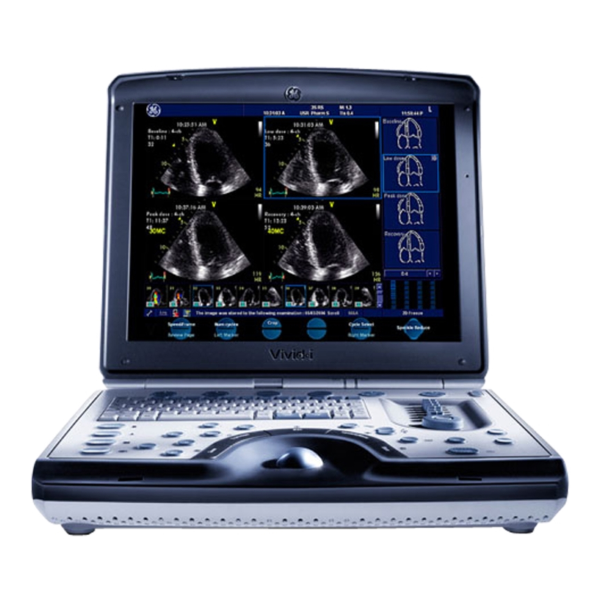
The GE VIVID I Ultrasound system is an advanced and versatile medical imaging technology designed to provide high-quality cardiac imaging for diagnostic purposes. Known for its portability, user-friendly interface, and exceptional imaging capabilities, the VIVID I Ultrasound system has become a valuable tool in various clinical settings for assessing cardiovascular health.
Key Features and Functionality:
1. Cardiac Imaging Excellence: The GE VIVID I Ultrasound system is specifically designed to deliver high-resolution images of the heart and its structures. It employs advanced transducer technology and imaging algorithms to provide detailed visuals of cardiac anatomy, function, and blood flow.
2. Portable Design: One of the standout features of the VIVID I is its portability. Unlike traditional ultrasound systems that are often bulky and stationary, the VIVID I is compact and lightweight. This makes it suitable for a wide range of clinical environments, including point-of-care situations, emergency departments, operating rooms, and outpatient settings.
3. User-Friendly Interface: The system features an intuitive user interface that facilitates ease of use. The user-friendly controls and menu options allow clinicians to quickly access imaging modes, adjust settings, and optimize imaging parameters.
4. Wide Range of Imaging Modes: The VIVID I offers a variety of imaging modes tailored to cardiac assessment. These include two-dimensional (2D) imaging for visualizing cardiac structures, Doppler imaging for assessing blood flow patterns, color flow imaging for detecting abnormalities in blood flow, and tissue Doppler imaging for evaluating tissue motion and strain.
5. Stress Echocardiography: The system is also equipped with stress echocardiography capabilities, allowing clinicians to assess the heart’s response to physical stress. This is particularly useful for diagnosing and monitoring heart conditions under exertion.
Clinical Applications:
The GE VIVID I Ultrasound system is widely used across various clinical applications within cardiology and related fields:
1. Echocardiography: The system’s exceptional imaging quality and versatile modes make it a powerful tool for echocardiography, enabling clinicians to visualize heart structures, evaluate valve function, assess myocardial performance, and detect cardiac abnormalities.
2.Cardiac Interventions: The real-time imaging capabilities of the VIVID I are beneficial during cardiac interventions. It provides visual guidance for procedures such as cardiac catheterization, pacemaker implantation, and other interventions requiring precise placement.
3. Emergency Medicine: In emergency situations, quick and accurate assessment of cardiac function is critical. The portability of the VIVID I makes it valuable for rapid bedside evaluations in emergency departments.
4. Outpatient Clinics: The system’s versatility and portability allow it to be utilized in outpatient settings, enabling clinicians to conduct routine cardiac assessments without the need for patients to visit specialized imaging centers.
Maintenance and Quality Assurance:
To ensure the optimal performance and reliability of the GE VIVID I Ultrasound system, regular maintenance is essential. Routine servicing, calibration, and software updates are recommended to maintain image quality and system functionality. Quality assurance programs, including periodic performance testing, help identify any deviations and ensure that the system consistently produces accurate and reliable results.
Conclusion:
The GE VIVID I Ultrasound system has transformed cardiac imaging by combining advanced technology with portability and user-friendliness. Its ability to provide high-quality cardiac visuals in various clinical settings has made it a cornerstone tool for cardiovascular assessment. Whether used in echocardiography, stress testing, emergency medicine, or outpatient clinics, the VIVID I system contributes to accurate diagnoses, informed treatment decisions, and improved patient care.
| Year Launched | 2004 |
| Estimated Market Price | Mid |
| Monitor | 15″ LCD |
| Monitor Resolution | |
| Image Size Resolution | 800*600 |
| Trackball or Trackpad | Trackball |
| CP Back-Lighting | Yes |
| Weight | 11lbs (5kg) |
| Probe Ports | 1 |
| Battery | O (up to 1hr) |
| Boot-Up Time | |
| Sleep Mode | |
| Maximum Depth of Field | 30cm |
| Minimum Depth of Field | 0-2cm |
| Cart | Yes(Option) |
| Independent Steer & Lockable Wheels | Yes |
| 2D, M mode | Yes |
| M-color Flow Mode | Yes |
| Anatomical M-mode | Yes |
| Color, Power Angio, Pulse Wave Doppler | Yes |
| SCW Doppler | Yes |
| Tissue Doppler(Velocity) Imaging | Yes |
| Stress Echo | Yes(Option) |
| Strain and Strain Rate | Yes(Option) |
| B Flow | Yes |
| Contrast Imaging-Cardiac | Yes(option) |
| Contrast Imaging – General Imaging | Yes |
| Tissue Harmonic Imaging | Yes |
| Speckle Reduction (=SRI) | Yes |
| Auto Image Opt(B mode) | Yes |
| Auto Image Opt(Doppler) | Yes |
| Triplex Mode | Yes |
| Auto IMT | Yes(Option) |
| Automated B/M/D Measurement | Yes |
| Automated LH Measurement(Automated Function Imaging(AFI), Cardiac Motion Quantification(CMQ), or Auto EF(Ejection Fraction) | Yes |
| Live Dual (B/BC) Mode | Yes |
| SmartExam or Scan Assistant | Yes |
| Raw Data File | Yes |
| Flexible Report | Yes |
| Abdominal | Yes |
| Women’s Health Care | Yes |
| OB | Yes |
| Vascular | Yes |
| TCD(Transcranial) | Yes |
| Small Parts | Yes |
| MSK/Anesthesiology | Yes |
| Pediatrics | Yes |
| Urology | Yes |
| Echocardiography_Adult | Yes |
| Echocardiography_Pediatric | Yes |
| Echocardiography_Neonate | Yes |
| Stress Echocardiography | Yes |
| Transesophageal Echo_Adult | Yes(Option) |
| Transesophageal Echo_Pediatric | Yes |
| Internal Medicine w/ Shared Service | Yes |
| Interventional Radiology | Yes |
| Contrast Imaging_Cardiac | Yes |
| Convex (1~6Mhz) | Yes |
| Biplane Micro Convex (3~10MHz) | |
| Linear (3~12Mhz) | Yes |
| Linear (<9Mhz) | Yes |
| Single Crystal Linear (3~12Mhz) | Yes |
| Single Crystal Linear (<9Mhz) | Yes |
| T or L shape Intra Operative | Yes |
| Phased Array_Adult | Yes |
| Phased Array_Pediatric | Yes |
| Phased Array_Neonate | Yes |
| ICE | Yes |
| TEE_Adult | Yes |
| TEE_Pediatric | Yes (9Mhz) |
| Pencil CW (2Mhz) | Yes |
| Pencil CW (5 or 6Mhz) | Yes |
| DICOM 3.0 | Yes |
| DICOM SR_Cardiac | Yes |
| DICOM SR_Vascular | Yes |
| DICOM SR_OB/GYN | Yes |
| JPEG, WMV, & AVI | Yes |
| USB | Yes(2) |
| HDD/SDD | 40GB |
| DVD/CD RW | Yes |
| Wireless LAN | Yes(Option) |


